Hydatidosis.
Echinococcosis, also known as hydatid disease, hydatid cyst, unilocular hydatid disease or cystic echinococcosis, is a potentially fatal parasitic disease that can affect many animals, including wildlife, commercial livestock and humans. The disease results from infection by tapeworm larvae of the genus Echinococcus - notably E. granulosus, E. multilocularis, E. vogeli and E. oligarthrus.
Infection cycle
Like many other parasite infections, the course of Echinococcus infection is complex. The worm has a life cycle that requires definitive hosts and intermediate hosts. Definitive hosts are normally carnivores such as dogs, while intermediate hosts are usually herbivores such as sheep and cattle. Humans function as accidental hosts, because they are usually a 'dead end' for the parasitic infection cycle.
The disease cycle begins with an adult tapeworm infecting the intestinal tract of the definitive host. The definitive host is usually a carnivore. The adult tapeworm then produces eggs which are expelled in the host's feces. Intermediate hosts, usually herbivores, become infected by ingesting the eggs of the parasite. Ingestion of eggs can occur by consumption of fecal contaminated food. Inside the intermediate host, the eggs hatch and release tiny hooked embryos (called oncospheres) which travel in the bloodstream, eventually lodging in an organ such as the liver, lungs and/or kidneys. There, they develop into hydatid cysts. Inside these cysts grow thousands of tapeworm larvae, the next stage in the life cycle of the parasite. When the intermediate host is predated or scavenged by the definitive host, the larvae are eaten and develop into adult tapeworms, and the infection cycle restarts.
Disease symptoms
As already noted, Echinococcus infection causes large cysts to develop in intermediate hosts. Disease symptoms arise as the cysts grow bigger and start eroding and/or putting pressure on blood vessels and organs. Large cysts can also cause shock if they happen to rupture.
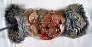
Infection with E. granulosus, common in "southern South America, the Mediterranean coast, the southern part of the former USSR, the Middle East, south-western Asia, northern Africa, Australia, New Zealand, Kenya and Uganda", typically results in the formation of hydatid cysts in the liver, lungs, kidney and spleen of the intermediate host. In echography or Computed tomography scans, hydatid cysts are often large with a flaky appearance (this is referred to as "hydatid sand"); this indicates the first stage of infection. In the second stage, medical imaging may show multiple daughter cysts. Hydatid cyst of liver can be accurately diagnosed by a serologic assay (Weinberg reaction, a specific example of complement fixation). However, the Weinberg reaction can be falsely negative; in one series, 38% of cases demonstrated a false negative result. Newer studies, such as ELISA, may be more sensitive. Eosinophilia is not a feature of cysts unless rupture occurs. In fact, usually there are no changes in blood biochemistry.
Hydatid disease of lung or liver is generally asymptomatic but can cause serious complications if rupture of the cyst occurs. Systemic anaphylaxis is usually associated with cyst rupture and can be predicted by positivity of Casoni reaction. There is also risk of intrapleural or intraperitoneal dissemination of the disease and of secondary infection that causes a lung or hepatic abscess. This condition is also known as cystic hydatid disease and can sometimes be successfully treated with surgery to remove the cysts. In Portugal there is also some experience with PAIR (Percutaneous Aspiration, Infusion of scolicidal agents and Reaspiration of cyst content) and medical therapy with albendazole alone in the dose of 400 mg twice daily. Therapy with albendazole or praziquantel should be initiated before any procedure and prolonged 28 days if dissemination of hydatid cyst is to be avoided.
Infection with E. multilocularis results in the formation of dense parasitic tumors in the liver, lungs, brain and other organs. Sometimes the infection in brain may cause tumour like symptoms and it needs removal by surgical means. This condition, also called alveolar hydatid disease is more likely to be fatal. Infection with Echinococcus vogeli, restricted to Central and South America is characterized by polycystic disease.
Unlike intermediate hosts, definitive hosts are usually not hurt very much by the infection. Sometimes, a lack of certain vitamins and minerals can be caused in the host by the very high demand of the parasite.
Diagnosis
Diagnosis of echinococcosis is made by using imaging and serology techniques. Cysts can be detected by ultrasonography, Computed tomography scans or occasionally by plain radiography. Detachment of cysts walls may lead to their collapse which gives rise to a radiological finding called "water lily sign". This finding is pathognomonic of echinococcosis.
The appearance can be similar to that of tumor metastasis.
Computed tomography
In the liver hydatid presents as a fluid density cyst, with frequent peripheral focal areas of calcification. Septation and daughter cysts may be visualised. Fluid is of variable density depending on the amount of proteinacous debris.
MRI
* T1 - mixed low signal (depending on the amount of proteinacous cellular debris) the walls and septae enhance.
* T2 - mixed high signal (depending on the amount of proteinacous cellular debris)
Prophylaxis
There are several strategies to prevent echinococcosis, most of which involve disruption of the parasite's life cycle. For instance, feeding raw offal to work dogs is a key point of infection in a farm environment and is strongly discouraged. Also, basic hygiene practices such as thoroughly cooking food and vigorous hand washing before meals can prevent the eggs entering the human digestive tract. Regular "deworming" of farm dogs with the drug praziquantel also helps kill the tapeworm. By employing such simple practices, hydatids have been virtually eliminated in New Zealand, where it was once very common. Effective vaccines, based on recombinant DNA technology, are being developed in Australia for sheep.
Investigations
* Blood CP
* Serology
* Casoni's Reaction
* X-Ray Abdomen
* USG and Computed tomography Scanning
* ERCP (Endoscopic retrograde Cholangio-Pancreatography)
Treatment
* Metronidazole 400-600 mg
* Albendazole
* Surgical
* Marsupialization
* Omentopexy
* Laminated Membrane Removal
* Mebendazole to prevent recurrence
Praziquan, tab 20 mg/kg 12 hourly for 2 weeks is given pre operatively
Surgery may be appropriate in certain cases.Fuentes:
http://en.wikipedia.org/wiki/Hydatidosis